

For pregnant ladies, ultrasounds are an informative (and typically vital) process. They sometimes produce two-dimensional black-and-white scans of fetuses that may reveal key insights, together with organic intercourse, approximate measurement, and abnormalities like coronary heart points or cleft lip. In case your physician desires a more in-depth look, they might use magnetic resonance imaging (MRI), which makes use of magnetic fields to seize pictures that may be mixed to create a 3D view of the fetus.
MRIs aren’t a catch-all, although; the 3D scans are tough for medical doctors to interpret properly sufficient to diagnose issues as a result of our visible system isn’t accustomed to processing 3D volumetric scans (in different phrases, a wrap-around look that additionally exhibits us the inside constructions of a topic). Enter machine studying, which might assist mannequin a fetus’s growth extra clearly and precisely from knowledge — though no such algorithm has been capable of mannequin their considerably random actions and numerous physique shapes.
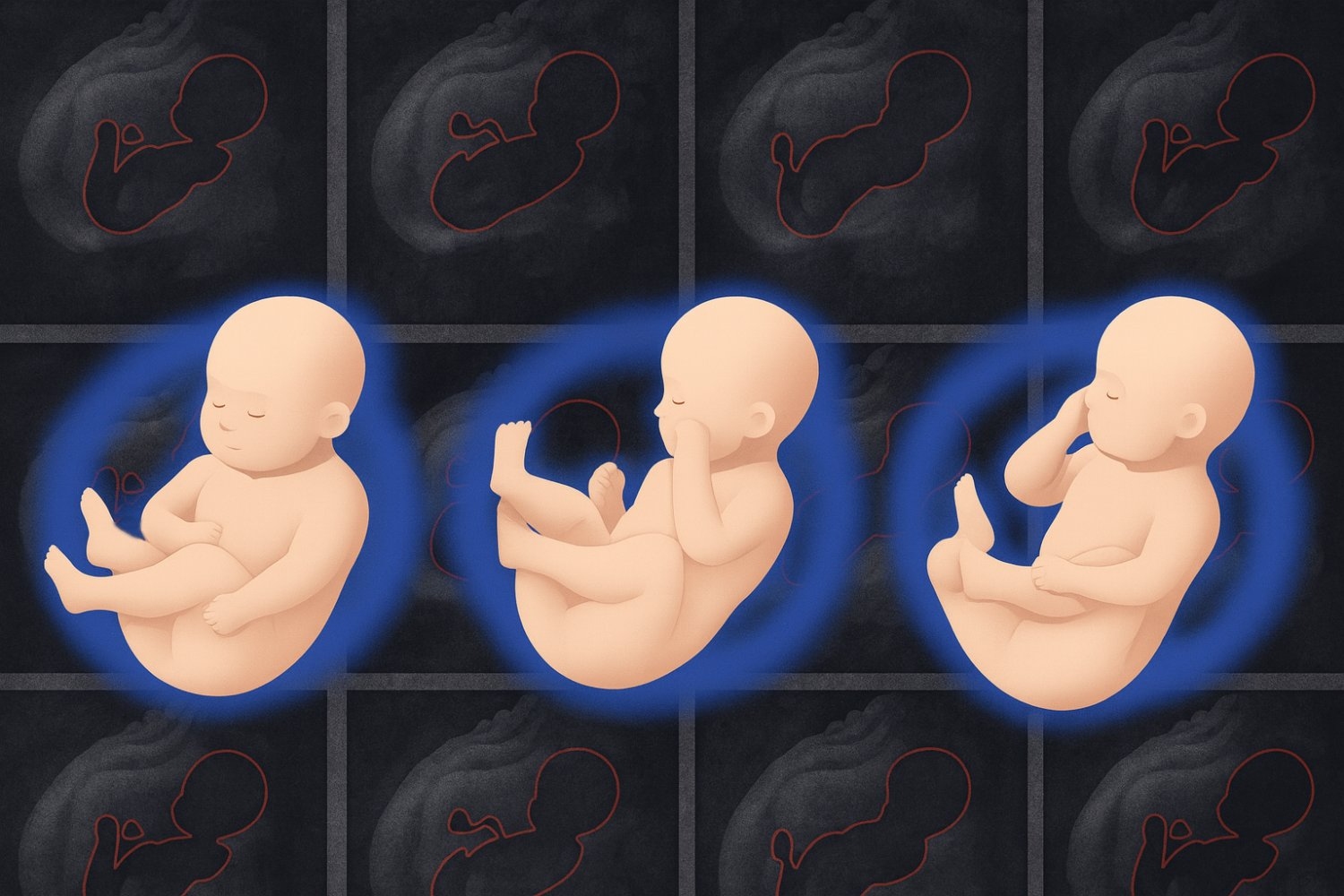
That’s, till a brand new method referred to as “Fetal SMPL” from MIT’s Pc Science and Synthetic Intelligence Laboratory (CSAIL), Boston Youngsters’s Hospital (BCH), and Harvard Medical College offered clinicians with a extra detailed image of fetal well being. It was tailored from “SMPL” (Skinned Multi-Particular person Linear mannequin), a 3D mannequin developed in laptop graphics to seize grownup physique shapes and poses, as a option to signify fetal physique shapes and poses precisely. Fetal SMPL was then skilled on 20,000 MRI volumes to foretell the situation and measurement of a fetus and create sculpture-like 3D representations. Inside every mannequin is a skeleton with 23 articulated joints referred to as a “kinematic tree,” which the system makes use of to pose and transfer just like the fetuses it noticed throughout coaching.
The in depth, real-world scans that Fetal SMPL realized from helped it develop pinpoint accuracy. Think about stepping right into a stranger’s footprint whereas blindfolded, and never solely does it match completely, however you appropriately guess what shoe they wore — equally, the device carefully matched the place and measurement of fetuses in MRI frames it hadn’t seen earlier than. Fetal SMPL was solely misaligned by a mean of about 3.1 millimeters, a niche smaller than a single grain of rice.
The method might allow medical doctors to exactly measure issues like the scale of a child’s head or stomach and examine these metrics with wholesome fetuses on the similar age. Fetal SMPL has demonstrated its medical potential in early assessments, the place it achieved correct alignment outcomes on a small group of real-world scans.
“It may be difficult to estimate the form and pose of a fetus as a result of they’re crammed into the tight confines of the uterus,” says lead writer, MIT PhD scholar, and CSAIL researcher Yingcheng Liu SM ’21. “Our method overcomes this problem utilizing a system of interconnected bones underneath the floor of the 3D mannequin, which signify the fetal physique and its motions realistically. Then, it depends on a coordinate descent algorithm to make a prediction, basically alternating between guessing pose and form from tough knowledge till it finds a dependable estimate.”
In utero
Fetal SMPL was examined on form and pose accuracy towards the closest baseline the researchers might discover: a system that fashions toddler development referred to as “SMIL.” Since infants out of the womb are bigger than fetuses, the crew shrank these fashions by 75 % to stage the enjoying subject.
The system outperformed this baseline on a dataset of fetal MRIs between the gestational ages of 24 and 37 weeks taken at Boston Youngsters’s Hospital. Fetal SMPL was capable of recreate actual scans extra exactly, as its fashions carefully lined up with actual MRIs.
The tactic was environment friendly at lining up their fashions to pictures, solely needing three iterations to reach at an affordable alignment. In an experiment that counted what number of incorrect guesses Fetal SMPL had made earlier than arriving at a remaining estimate, its accuracy plateaued from the fourth step onward.
The researchers have simply begun testing their system in the actual world, the place it produced equally correct fashions in preliminary medical assessments. Whereas these outcomes are promising, the crew notes that they’ll want to use their outcomes to bigger populations, completely different gestational ages, and a wide range of illness circumstances to higher perceive the system’s capabilities.
Solely pores and skin deep
Liu additionally notes that their system solely helps analyze what medical doctors can see on the floor of a fetus, since solely bone-like constructions lie beneath the pores and skin of the fashions. To raised monitor infants’ inside well being, comparable to liver, lung, and muscle growth, the crew intends to make their device volumetric, modeling the fetus’s inside anatomy from scans. Such upgrades would make the fashions extra human-like, however the present model of Fetal SMPL already presents a exact (and distinctive) improve to 3D fetal well being evaluation.
“This examine introduces a way particularly designed for fetal MRI that successfully captures fetal actions, enhancing the evaluation of fetal growth and well being,” says Kiho Im, Harvard Medical College affiliate professor of pediatrics and employees scientist within the Division of New child Drugs at BCH’s Fetal-Neonatal Neuroimaging and Developmental Science Heart. Im, who was not concerned with the paper, provides that this method “is not going to solely enhance the diagnostic utility of fetal MRI, but additionally present insights into the early useful growth of the fetal mind in relation to physique actions.”
“This work reaches a pioneering milestone by extending parametric floor human physique fashions for the earliest shapes of human life: fetuses,” says Sergi Pujades, an affiliate professor at College Grenoble Alpes, who wasn’t concerned within the analysis. “It permits us to detangle the form and movement of a human, which has already confirmed to be key in understanding how grownup physique form pertains to metabolic circumstances and the way toddler movement pertains to neurodevelopmental problems. As well as, the truth that the fetal mannequin stems from, and is appropriate with, the grownup (SMPL) and toddler (SMIL) physique fashions, will enable us to check human form and pose evolution over lengthy durations of time. That is an unprecedented alternative to additional quantify how human form development and movement are affected by completely different circumstances.”
Liu wrote the paper with three CSAIL members: Peiqi Wang SM ’22, PhD ’25; MIT PhD scholar Sebastian Diaz; and senior writer Polina Golland, the Sunlin and Priscilla Chou Professor of Electrical Engineering and Pc Science, a principal investigator in MIT CSAIL, and the chief of the Medical Imaginative and prescient Group. BCH assistant professor of pediatrics Esra Abaci Turk, Inria researcher Benjamin Billot, and Harvard Medical College professor of pediatrics and professor of radiology Patricia Ellen Grant are additionally authors on the paper. This work was supported, partially, by the Nationwide Institutes of Well being and the MIT CSAIL-Wistron Program.
The researchers will current their work on the Worldwide Convention on Medical Picture Computing and Pc Assisted Intervention (MICCAI) in September.